Who Knew WashU?
Question: The Record has served as the go-to news source for the WashU community for many years. It began as a weekly printed newspaper but transitioned to a digital publication in 2010. When was the first issue?
Answer: B) 1974. This year marks the Record’s 50th anniversary of bringing the campus community news about the many accomplishments of our faculty, staff and students.
Congrats to this week’s winner, Kweku Enninful, a graduate student at the McKelvey School of Engineering who will receive an “I Knew WashU” luggage tag!
Campus and community news
Notables
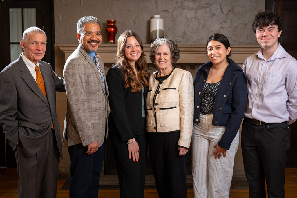
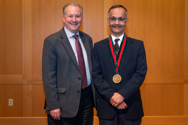
Ethic of Service Award honors investment in St. Louis
Each year, the Gerry and Bob Virgil Ethic of Service Award recognizes select members of the WashU community who show exemplary commitment to service and engagement within the St. Louis area.

Subscribe to the Record
Videos
The hidden river
The Mississippi River defines St. Louis, shaping its life and culture. But today, for many St. Louisans, that connection has been broken, says Derek Hoeferlin, chair of landscape architecture in the Sam Fox School of Design & Visual Arts at Washington University in St. Louis.